South Australian Medical Heritage Society Inc
Website for the Virtual Museum
Home
Coming meetings
Past meetings
About the Society
Main Galleries
Medicine
Surgery
Anaesthesia
X-rays
Hospitals,other organisations
Individuals of note
Small Galleries
Ethnic medicine
- Aboriginal
- Chinese
- Mediterran
Old x-ray equipment in South Australia
Acknowledgments: We are grateful to Dr Bernard Vaughan who kindly provided the following text and photographs. We are also grateful to the Department of Physics at the University of Adelaide and to David Buob, president of the Glenside Historical Society who allowed us to photograph some of the items .
The news of the discovery of x-rays by Professor Conrad Roentgen in December 1895 reached the colony of South Australia when the following cable from London was published in the “South Australian Register”.
London 30 Jan. 1986. “Great interest is being excited by experiments in the application to medical purposes of Roentgen’s newly discovered method of photographing through opaque substances. It seems the results of the most astounding character have been obtainable. Gall stones and bladder stones have been detected , and injuries to the bones easily seen through the flesh”.
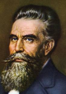
Professor Wilheim Conrad Roentgen
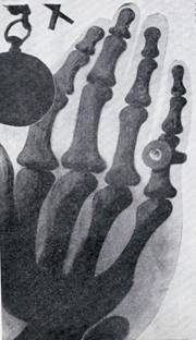
X-ray by William Bragg of a hand May 1886
(Two pictures from “X.Ray Pioneers of Australia” published By Watson & Sons Ltd 1946)
William Bragg, professor of Physics at Adelaide University, was the first South Australian to repeat Professor Roentgen’s experiments. At first he used Crooks Tubes made by Mr AL Rogers, his laboratory assistant, and later in May used tubes manufactured by Reynolds and Branson of Leeds UK. These had been brought back by Mr S Barbour, a local business man representing FW Faulding & Company who happened to be in England earlier in the year.
On May 28th. using Barbour’s tubes and a 12” Ruhmkorff coil Professor Bragg succeeded in obtaining an x-ray photograph of his hand which was published in the “Australasian”. On June 17th Professor Bragg delivered a public lecture & demonstration.
Sir William Bragg, and his son Sir Lawrence Bragg were jointly awarded the Nobel Prize for their research in X. Ray crystallography in 1915. An exhibition of Professor William Bragg’s apparatus is to be set up in the Physics department of Adelaide University in 2006. The following photographs of early X.Ray tubes which are stored in the Physics department of Adelaide University[[ to be inserted - Public display and lecture on x.rays , Painting by R.A.Thomas]]
The following illustrations are of early X-Ray tubes kept in the department of Physics of Adelaide University. Until 1913 the tubes were evacuated to less than a millionth of an atmosphere, and contained a cathode and an anode. The electrons in the cathode beam were derived from the remaining gas which became ionised. X-Rays were produced when the accelerated electrons struck the anode. The electricity was provided by a battery energising a “Ruhmkorff Coil” with a mercury “make and break” powered by the magnetism produced by the low tension windings, the rapidly fluctuating current inducing a very high voltage in the high tension windings. The tubes “Crook’s Tubes” were named after the inventor.
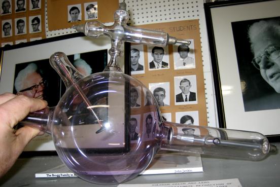
A Crooks x-ray tube used by Professor William Bragg in his experiments
Crook’s tubes were erratic in behaviour, and the only adjustment was the duration of the exposure. In 1913 William Coolidge working for “General Electric” in the U.S.A. designed an X-Ray tube which is still the principle of the modern X-Ray tube. It was completely evacuated and the electrons from the cathode were produced by an electrically heated filament, so that the current could be regulated. The cathode rays were focussed on to the anode which consisted of a tungsten target embedded in a large piece of copper. The production of X-Rays is a very inefficient process, with a lot of heat produced. The high tension voltage is now produced by a transformer, with the low tension voltage derived from the electric mains via an autotransformer, allowing selection of the final Kilo-voltage. (60-110 KV.)
The circuit was self rectifying, but a diode valve was incorporated to prevent the reverse current burning out the cathode. Modern Coolidge tubes have a rotating anode which distributes the heat around the edge of a disc rotated by an induction motor, The high tension voltage is now fully rectified.
An early Coolidge tube preserved in the Physics Department of Adelaide University. Note cathode filament, with focussing shield & a heavy anode.
A collection of old x-ray tubes in Adelaide University Physics Department.
There is a Coolidge tube with an anode heat sink in the front on the right.
Behind on the right is a Crook’s tube with additional anodes to deflect the cathode rays on to the main copper anode, a practice which was discontinued. The tube on the left is a Coolidge tube with a heavy anode, and a heat finned sink to assist cooling.
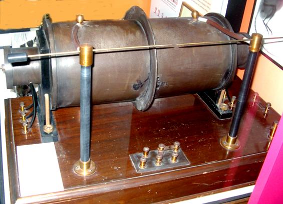
The Ruhmkorff coil used by William Bragg in early x ray experiments
Heinrich Ruhmkorff (1803-1877) invented an induction coil that could produce sparks more than a foot in length. This coil was used in the first radio transmitters and also to energise Crook’s tubes for early x-ray experiments.
Now follow photographs of preserved early x-ray apparatus at present stored in the museum section of Glenside Hospital
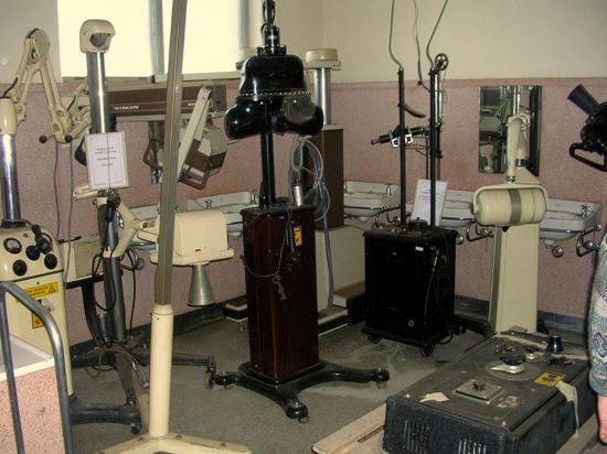
Collection of old x-ray equipment stored at Glenside Hospital
The generator & control of an “Elgen” mobile x-ray machine 1920.
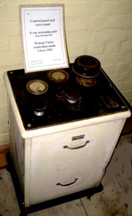
1920 Watson Victor generator
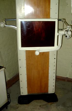
and screening stand with fluoroscope (below)
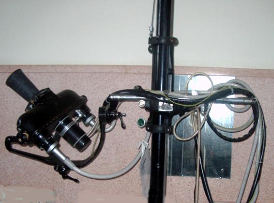
Phillips Stanford mobile 1960
Note light beam diaphragm
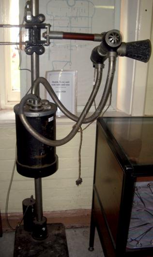
Citca mobile x-ray machine 1920
Note cooling vents for Coolidge tube anode
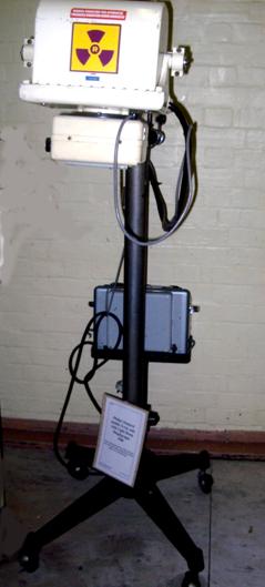
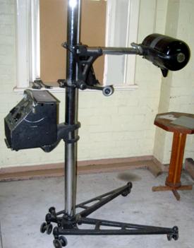
Watson-Victor S.F.I.mobile unit 1951
An old x-ray tube housing with a cone mounted on an adjustable arm. It was free to move up and down on a column, which was free to move on floor and ceiling rails
A non-shockproof dental unit circa 1920
-o0o-